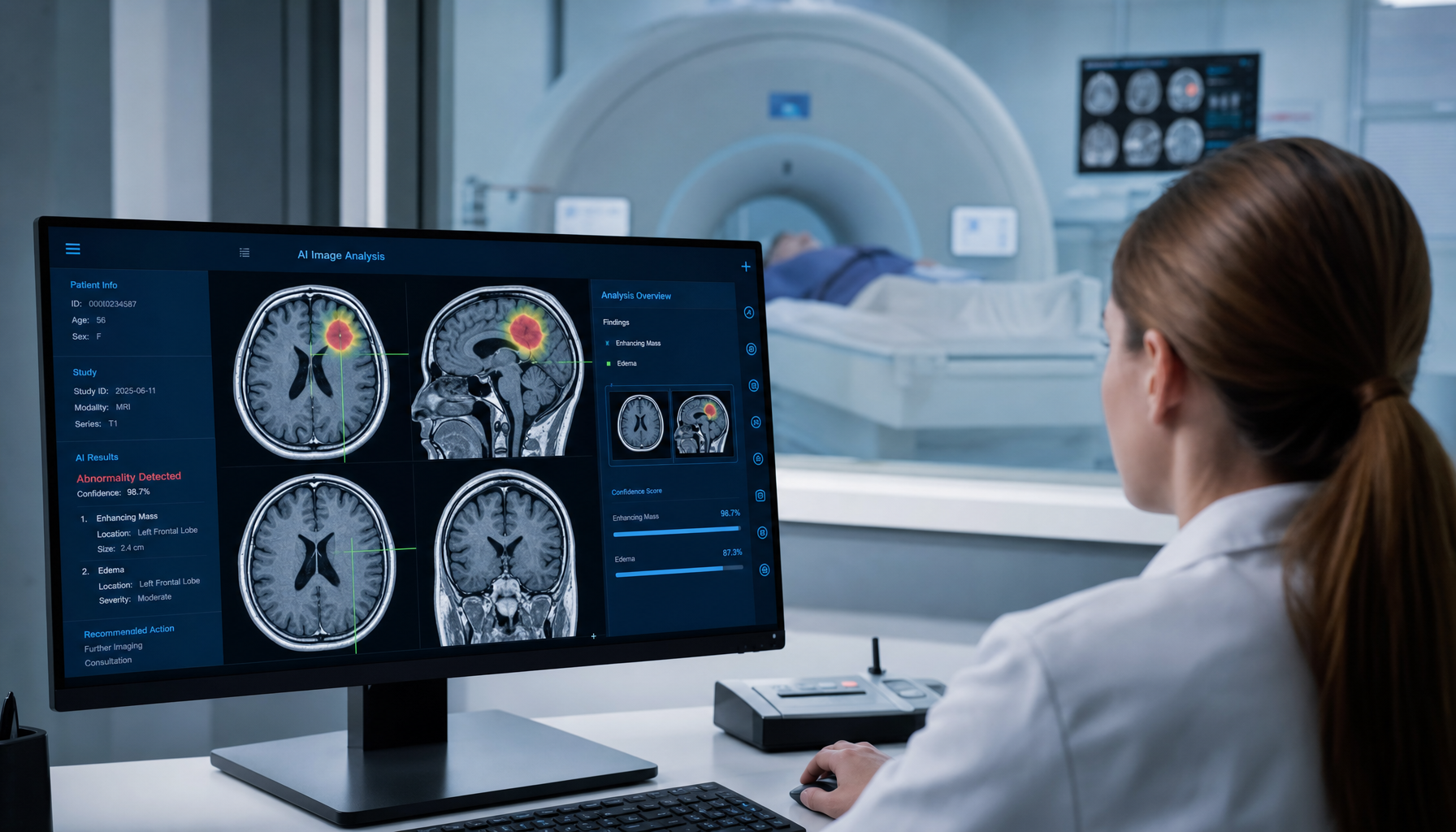
AI Radiology Diagnostic Systems Surpass Human Physicians: FDA Approves Autonomous Interpretation Platform
Deep learning models exceed radiologist accuracy in CT and MRI interpretation
AI medical imaging diagnosis crossed a critical threshold in 2025-2026 — upgrading from "assistive tool" to "autonomous interpretation system" and receiving the FDA's first autonomous approval decision authority.
FDA Milestone
In October 2025, the FDA granted De Novo classification approval to the first AI diagnostic system requiring no human radiologist review — Annalise.ai's Annalise CT Brain platform, capable of autonomously interpreting non-contrast head CT scans in acute stroke patients, detecting intracranial hemorrhage, large vessel occlusion, and early ischemic changes, and generating complete structured reports.
In December of the same year, Zebra Medical Vision (now renamed Nanox AI) received autonomous interpretation approval for its HealthGATE chest X-ray platform, capable of detecting 11 common pathologies (including lung nodules, pneumothorax, pleural effusion, cardiomegaly), achieving AUC of 0.94-0.97 on a 140,000-image validation set — outperforming radiology residents (AUC 0.89-0.92).
Key Clinical Validation
Head-to-Head Comparison Studies: AI vs. Radiologists (2025-2026):
- Lung nodule detection (LIDC-IDRI dataset + real-world retrospective): AI (n = 5 models) average sensitivity 94.7%, radiologists average 88.3%; false-positive rate 1.2/scan vs. 2.5/scan
- Breast cancer screening (Swedish Mammography Screening Trial): AI-assisted screening cancer detection rate 6.1/1,000, traditional double reading 5.3/1,000; AI group recall rate reduced by 37%
- Head CT intracranial hemorrhage (MARS study, n = 7,000): AI negative predictive value reached 99.8%, enabling safe hemorrhage exclusion and eliminating emergency physicians' waiting time for radiology reports
Transforming Clinical Workflow
AI integration is redesigning radiology workflow:
- Pre-screening queue: AI prioritizes abnormal images (intracranial hemorrhage, pneumothorax, pulmonary embolism), reducing emergency report waiting time from an average of 45 minutes to 8 minutes
- Quantitative analysis: AI not only detects lesions but also automatically performs measurements (nodule volume, stroke core area, bone density T-score), eliminating human measurement variability
- Structured reporting: AI automatically generates structured report drafts meeting ACR standards, requiring only radiologist review and signature — a 2025 Mayo Clinic pilot showed a 65% reduction in report writing time
Market and Challenges
The global AI medical imaging market is projected to grow from $3.5 billion in 2025 to $12 billion by 2030. However, AI autonomous interpretation still faces major challenges: data distribution differences across institutions reducing model generalization, legal liability attribution (who bears responsibility for AI decision errors), and radiologists' resistance to being "replaced." The American College of Radiology (ACR)'s position is that AI is a "collaborative partner" rather than a replacement, with human radiologists always ultimately responsible for final reports.
Future Directions
Multimodal AI (integrating CT, MRI, PET, pathology, and genetic data for comprehensive diagnosis), federated learning (multi-center data training without sharing patient privacy), and real-time AI (interpreting intraoperative images in real time within the operating room) will be the most impactful development directions in the next three years.
POC.HK Future Technology Observatory — Independent Technology Watch Report